
Articles
- Page Path
- HOME > J Korean Powder Metall Inst > Volume 28(4); 2021 > Article
-
Article
- Fabrication of Layered Cu-Fe-Cu Structure by Cold Consolidation of Powders using High-pressure Torsion
- Peyman Asghari-Rada,b, Yeon Taek Choia, Nhung Thi-Cam Nguyena,b, Praveen Sathiyamoorthia,b, Hyoung Seop Kima,b,c,d,*
-
Journal of Korean Powder Metallurgy Institute 2021;28(4):287-292.
DOI: https://doi.org/10.4150/KPMI.2021.28.4.287
Published online: July 31, 2021
a Department of Materials Science and Engineering, Pohang University of Science and Technology (POSTECH), Pohang, 37673, Republic of Korea
b Center for High Entropy Alloys, Pohang University of Science and Technology (POSTECH), Pohang 37673, Republic of Korea
c Graduate Institute of Ferrous Technology (GIFT), Pohang University of Science and Technology (POSTECH), Pohang 37673, Republic of Korea
d Institute for Materials Research, Tohoku University, Sendai 980-8577, Japan
- *Corresponding Author: Hyoung Seop Kim, TEL: +82-54-279-2150, FAX: +82-54-279-2399, E-mail: hskim@postech.ac.kr
- - P. Asghari-Rad·Y. T.Choi·N. Thi-Cam Nguyen: 학생, P. Sathiyamoorthi·H. S. Kim: 교수
© The Korean Powder Metallurgy Institute. All rights reserved.
- 1,873 Views
- 8 Download
- 2 Crossref
Abstract
- In this study, the layered structures of immiscible Fe and Cu metals were employed to investigate the interface evolution through solid-state mixing. The pure Fe and Cu powders were cold-consolidated by high-pressure torsion (HPT) to fabricate a layered Cu-Fe-Cu structure. The microstructural evolutions and flow of immiscible Fe and Cu metals were investigated following different iterations of HPT processing. The results indicate that the HPTprocessed sample following four iterations showed a sharp chemical boundary between the Fe and Cu layers. In addition, the Cu powders exhibited perfect consolidation through HPT processing. However, the Fe layer contained many microcracks. After 20 iterations of HPT, the shear strain generated by HPT produced interface instability, which caused the initial layered structure to disappear.
- Severe Plastic Deformation (SPD) is an illustrious technique to produce ultrafine/nano-grained materials by inducing large plastic strain [1, 2]. Among the SPD techniques, high-pressure torsion (HPT) is one of the most attractive methods in processing materials with exceptional grain refinement by simultaneous application of high hydrostatic pressure and torsional shear strain [3-5]. Besides grain refinement, HPT has been utilized to bond machining chips and consolidate metallic powders with uttermost densification [6-9].
- Several comprehensive investigations have been done on employing the HPT technique to achieve direct diffusion bonding between two metals/alloys such as Al-Mg, Al-Cu, and Cu-Mo systems [10-12]. Accordingly, the HPT processing has been demonstrated to promote the solid-state reactions by intensive rapid diffusion due to HPT-induced lattice defects, including vacancies, dislocation, and grain boundaries [11]. However, investigation on solid-state reactivity and interface evolution of the metals with immiscible nature through SPD processing are limited [13, 14]. Bachmaier et al. conducted two-step SPD processing on immiscible Fe-Cu system and fabricated supersaturated solid solutions of Fe-Cu alloy with structures far-from-equilibrium state [13]. Also, Sauvage et al. subjected the Fe-Cu system to a cold drawing process followed by HPT and revealed a significant interdiffusion of Cu and Fe, leading to the fabrication of a nonequilibrium supersaturated solid solution structure [14].
- In the present study, we demonstrate the fabrication of a layered Cu-Fe-Cu structure through cold-consolidation of pure Fe and Cu powders utilizing HPT processing. Further, systematic microstructural evolution at the interface between the Fe and Cu layers at different HPT turns was investigated.
1. Introduction
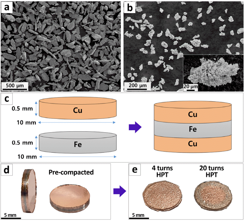
- The as-received pure Fe and Cu powders were used as starting materials in the present study. Micrographs of the pure Fe and Cu powder particles are illustrated in Fig. 1a and 1b, respectively. Both the powders exhibit large particle sizes with irregular morphology. The Fe powders have an average particle size of ~200 μm, and the electrolytic Cu powders consist of fine particles with an average size of ~3 μm, as shown in Fig. 1b. To fabricate the layered Cu-Fe-Cu samples, the following procedure was performed. First, the Fe and Cu powders were pre-compacted separately into a disk shape (ϕ1 0 mm × 0.5 mm) under a pressure of 20 MPa using a hand-press machine (Fig. 1c). Then, the disk samples were arranged in a sequence of Cu-Fe-Cu and compacted under the pressure of 40 MPa using the hand-press machine (Fig. 1d). Figure 1c illustrates the schematic of the pre-compacted Cu- Fe-Cu layered structure. At the last stage, the layered pre-compacted samples were consolidated using the HPT process under a pressure of 6 GPa, a rotation rate of 1r pm, and a rotation number of 4 and 20. Figure 1e shows the HPT-processed bulk samples after 4 and 20 turns.
- An optical microscope (OM) and a field emission scanning electron microscope (Fe-SEM) equipped with backscattered electron (BSE) and energy-dispersive X-ray spectroscopy (EDS) detectors were performed to analyze the microstructural evolution after different turns of HPT processing. The specimens for microstructural analyses were prepared by mechanical polishing using SiC papers up to 1200 grit, followed by fine polishing using diamond paste (3 μm and 1μm ) and colloidal silica.
2. Experimental methods and procedure
SEM micrographs of pure (a) Fe and (b) Cu powders, (c) schematic illustration of the layered structure fabrication through pre-compaction of Fe and Cu powders, (d) image of the pre-compacted layered samples, and (e) image of HPTprocessed samples after 4 and 20 turns.
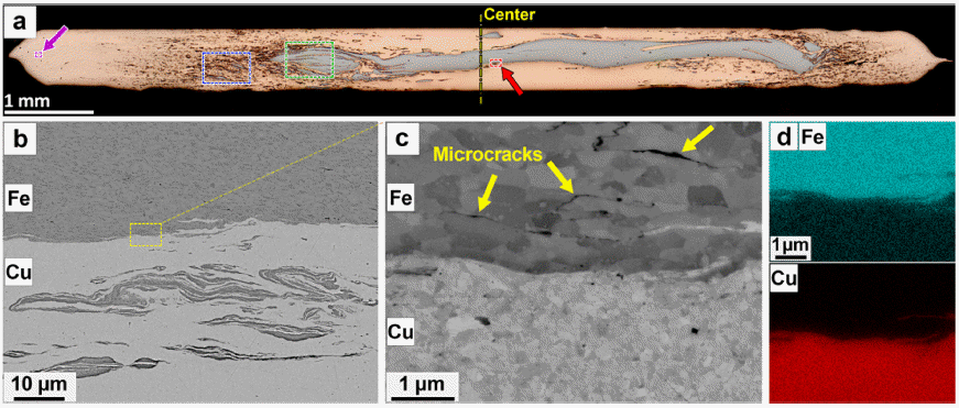
- Figure 2a shows the OM micrograph of the cross-sectional area of the four turns processed HPT sample. The top and bottom layers (orange color) correspond to the Cu layers, and the inner layer (gray color) corresponds to the Fe layer. Figure 2b shows the BSE micrograph taken from the area marked with a yellow dashed square in Fig. 2a. Also, the corresponding EDS maps displaying the elemental distribution are illustrated in Fig. 2c.
- Generally, the powder consolidation through HPT processing can be explained by the following mechanism [15-17]. First, the deformation of powders and the closure of pores between the powders occur to some extent under the applied hydrostatic pressure (6 GPa pressure in the present study). Under the application of torsional strain, powders are subjected to large shear strain, which can significantly deform and refine the powders and close the pores further. Accordingly, the samples fabricated by HPT exhibit ultra-high densification compared to the samples fabricated by other powder metallurgybased techniques [7, 8]. However, the formability of powders is another parameter that controls achieving a fully dense structure. After four turns of HPT processing in the present study, the Cu layers demonstrate almost perfect consolidation, while many cracks appear in the inner Fe layer (Fig. 2a, 2b). The compressibility of the Cu powder could be due to its crystalline structure (facecantered cubic (FCC)), which leads to a proper consolidation during HPT processing. The Fe powders with body-centered cubic (BCC) structure do not show an appropriate response under the shear strain, resulting in crack formation in the Fe layer (Fig. 2b). Furthermore, a close look at the interface of Fe and Cu layers reveals a drastic change in the chemical composition between these two pure metals (Fig. 2c). It seems that the Fe and Cu layers just formed a physical contact and there is no evidence for the formation of chemical compounds at the interface.
- An OM micrograph taken from the cross-sectional area of the HPT-processed sample for twenty turns is shown in Fig. 3a. According to this figure, in the central region of the disk, the inner Fe layer keeps its continuity, although the interfaces of the Fe and Cu layers are no longer smooth. Conversely, the continuity of the inner Fe layer is eliminated after twenty turns of HPT at the outer parts of the disc-shaped sample. It is noteworthy that the amount of shear strain created during HPT is proportional to the distance from the center of the disk, so that the greater the distance from the center of the disk, the greater the amount of shear strain applied [18, 19]. Consequently, the observed evolution of the inner Fe layer results from interface instability generated by the shear strain during the solid-state mixing.
- The high-magnification image of an area in the central region of the disk (indicated by an arrow and a red dashed square in Fig. 3a) is displayed in Fig. 3b. This image clearly shows the instability of the interface between two immiscible metals after solid-state mixing. The smooth interface transforms to an irregular wavy interface, and also part of the Fe layer is fragmented and penetrated toward the Cu layer, forming a composite-like structure. Figure 3c presents the area marked by a yellow dashed square in Fig. 3b. In this figure, Fe and Cu grains are apparent on both sides of the interface, and observations indicate that the average grain size in the Cu layer is about 300 nm which is finer than that in the Fe layer (about 700 nm). Moreover, a closer look reveals numerous intergranular microcracks in the Fe layer; however, no crack is observed in the Cu layer. The formation of these microcracks under the shear strain in the Fe layer could result in the rupture of pieces from the Fe layer, and eventually, these separated pieces move towards the Cu layer.
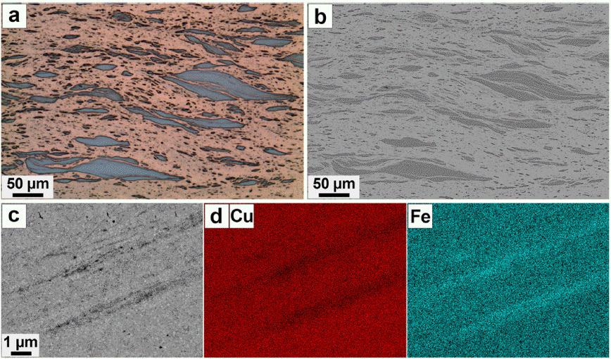
- Figures 4a and 4b show the microstructure analysis of the twenty-turns HPT-processed sample at a midway position between the center and edge, marked with a green dashed square in Fig. 3a. The OM and corresponding BSE micrographs present the fragmentation of the Fe layer and the formation of a fine laminate Fe-Cu structure. The difference in microstructural observation compared to the center position can be attributed to higher shear strain. A high magnification image of the area indicated by a yellow dashed square in Fig. 4b is displayed in Fig. 4c. The Cu strips that are spread between the fragmented Fe layers can be seen evidently in Fig. 4c. An EDS line scanning is conducted through these strips along the yellow dashed line, as indicated in Fig 4c. The EDS results (Fig. 4d) indicate that there is no dissolution of Cu in the Fe region, and the chemical composition change at the interface is almost sharp. However, the composition of the Cu strips presents a low content of the Fe element. Since the thickness of the Cu strip is narrow and the EDS analysis collects the data from a vast subsurface region, the presence of a low Fe content in the Cu strips could be attributed to the limitation of EDS analysis be ignored.
- The area marked with a blue dashed square in Fig. 3a is shown in Fig. 5a and 5b. As can be seen in the OM and BSE images, the continuity of the Fe layer is eliminated, and the islands of Fe particles are dispersed through the Cu matrix. Figure 5c presents an area at the edge of the HPT disk marked by a purple arrow and a dashed square in Fig. 3a. Also, the corresponding EDS maps showing the distribution of Cu and Fe elements are represented in Fig. 5d. Since the edge part of the disk experiences high shear strain, intensive mixing of Fe and Cu elements occurs, becoming difficult to distinguish the Fe and Cu interface by SEM-EDS analysis.
- The Fe-Cu system is immiscible at room temperature due to the positive enthalpy of mixing [20]. However, HPT processing can extend the mutual solubility of Fe and Cu metals due to the introduction of intensive lattice defects, leading to the formation of non-equilibrium supersaturated solid solutions [13, 14]. In the present study, the alloying of Fe and Cu could happen at the edge region of the HPT disk (Fig. 5c and 5d); however, further microstructural analysis using atom-probe tomography (APT) is necessary for validation of Fe-Cu alloying that it will be the subject for our future work.
3. Results and Discussion
(a) OM micrograph taken from the cross-sectional area of the sample HPT-processed for four turns, (b) SEM-BSE image taken from the region marked by a yellow dashed square in section (a), and (c) corresponding SEM-EDS maps of Cu and Fe elements.

(a) OM micrograph taken from the cross-sectional area of the sample HPT-processed for twenty turns, (b) SEM-BSE image taken from the central region of the HPT disk marked by a red arrow and dashed square in section (a), (c) SEM-BSE image from the region marked by a yellow dashed square in section (b), and (d) corresponding SEM-EDS maps of Cu and Fe elements.
(a) OM micrograph taken from the area marked by green dashed square in Fig. 3(a), (b) corresponding SEM-BSE image of section (a), (c) magnified SEM-BSE image from the region marked by a yellow dashed square in section (a, b), and (d) EDS line-scanning taken from along the dashed like depicted in section (c).

(a) OM micrograph taken from the area marked by a blue dashed square in Fig. 3(a), (b) corresponding SEM-BSE image of section (a), (c) SEM-BSE image taken from the region marked by a purple arrow in Fig. 3(a), and (d) corresponding SEM-EDS maps of Cu and Fe elements.
- In the current study, the layered Cu-Fe-Cu structure was fabricated by consolidating pure Fe and Cu powders using the HPT process. The Cu layer demonstrated a fully dense structure after HPT; however, the Fe layer contained many microcracks. The interface stability between Fe and Cu layers was investigated through different four and twenty turns of HPT processing. The immiscible Fe and Cu metals demonstrated a sharp chemical boundary after four turns of HPT. However, the initial layered structure was eliminated after twenty turns of HPT processing due to the interface instability caused by shear strain.
4. Conclusion
-
Acknowledgements
- This research was financially supported by the National Research Foundation of Korea (NRF: 2021R1A2C3006662) and the Ministry of Trade, Industry, and Energy (20000495).
Acknowledgments
- 1. R. N. Harsha, V. M. Kulkarni and B. S. Babu: Mater. Today Proc., 5 (2018) 22340..Article
- 2. G. Faraji and H. S. Kim: Mater. Sci. Tech., 33 (2017) 905..Article
- 3. A. P. Zhilyaev and T. G. Langdon: Prog. Mater. Sci., 53 (2008) 893..Article
- 4. N. T. C. Nguyen, P. Asghari-Rad, P. Sathiyamoorthi, A. Zargaran, C. S. L ee a nd H . S. K im: Nat. Commun., 11 (2020) 2736..ArticlePubMedPMC
- 5. S. Hadi, F. R. Lotfabad, M. H. Paydar and R. Ebrahimi: Met. Mater. Int., (2020) (in press)..
- 6. K. Edalati, Y. Yokoyama and Z. Horita: Mater. Trans., (2010) 0911300948-0911300948..
- 7. P. Asghari-Rad, P. Sathiyamoorthi, N. T. C. Nguyen, A. Zargaran, T. S. Kim and H. S. Kim: Scr. Mater., 190 (2021) 69..Article
- 8. P. Asghari-Rad, N. T. C. Nguyen, Y. Kim, A. Zargaran, P. Sathiyamoorthi and H. S. Kim: Mater. Lett., 303 (2021) 130503..Article
- 9. S. D. Oguntuyi, O. T. Johnson and M. B. Shongwe: Met. Mater. Int., 27 (2021) 2146..Article
- 10. X. Qiao, X. Li, X. Zhang, Y. Chen, M. Zheng, I. S. Golovin, N. Gao and M. J. Starink: Mater. Lett., 181 (2016) 187..Article
- 11. K. Oh-Ishi, K. Edalati, H. S. Kim, K. Hono and Z. Horita: Acta Mater., 61 (2013) 3482..Article
- 12. V. Tavakkoli, A. Mazilkin, T. Scherer, M. Mail, Y. Beygelzimer, B. Baretzky, Y. Estrin and R. Kulagin: Mater. Lett., 302 (2021) 130378..Article
- 13. A. Bachmaier, M. Kerber, D. Setman and R. Pippan: Acta Mater., 60 (2012) 860..ArticlePubMedPMC
- 14. X. Sauvage and R. Pippan: Mater. Sci. Eng. A, 410 (2005) 345..Article
- 15. K. Edalati, Z. Horita, H. Fujiwara and K. Ameyama: Metall. Mater. Trans. A, 41 (2010) 3308..Article
- 16. J. M. Cubero-Sesin and Z. Horita: Mater. Sci. Eng. A, 558 (2012) 462..Article
- 17. Y. J. Zhao, R. Massion, T. Grosdidier and L. S. Toth: IOP Conf. Ser. Mater. Sci. Eng., 63 (2014) 012032..Article
- 18. R. Valiev: Acta Mater., 44 (1996) 4705..Article
- 19. P. Asghari-Rad, P. Sathiyamoorthi, J. W. Bae, H. Shahmir, A. Zargaran and H. S. Kim: Adv. Eng. Mater., 22 (2020) 1900587..Article
- 20. A. R. Yavari, P. J. Desre and T. Benameur: Phys. Rev. Lett., 68 (1992) 2235..ArticlePubMed
Figure & Data
References
Citations
- Characterization of Cu-5Fe (wt.%) fabricated by powder consolidation using high-pressure torsion
Amar Djemli, Hiba Azzeddine, Piotr Bazarnik, Foudil Sahnoune, Yi Huang, Thierry Baudin, François Brisset, Megumi Kawasaki, Terence G. Langdon
Journal of Materials Science.2025; 60(40): 19267. CrossRef - Supreme tensile properties in precipitation-hardened 316L stainless steel fabricated through powder cold-consolidation and annealing
Do Won Lee, Peyman Asghari-Rad, Yoon-Uk Heo, Sujung Son, Hyojin Park, Ji-Su Lee, Jae-il Jang, Byeong-Joo Lee, Hyoung Seop Kim
Materials Science and Engineering: A.2024; 893: 146107. CrossRef
 ePub Link
 Cite this Article
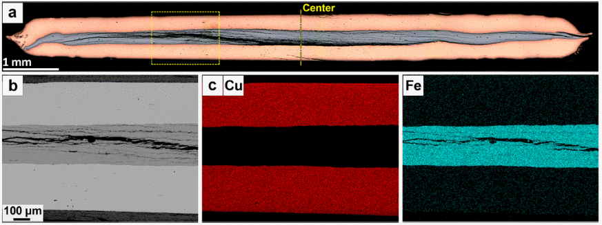

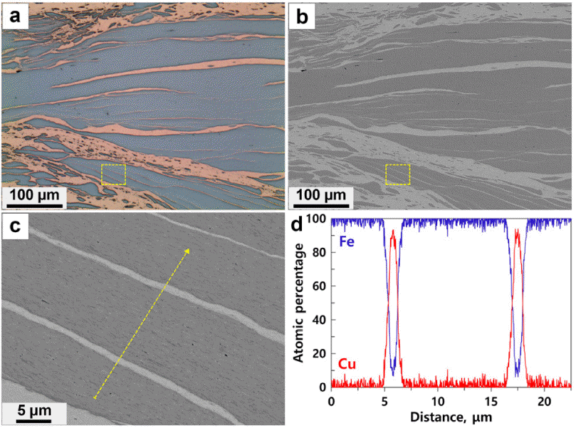


Fig. 1
Fig. 2
Fig. 3
Fig. 4
Fig. 5
TOP
