Articles
- Page Path
- HOME > J Korean Powder Metall Inst > Volume 27(6); 2020 > Article
-
ARTICLE
- Features of Nickel Nanoparticles Structure Synthesized by the Spark Discharge Method
- C. K. Rheea,*, A. D. Maksimovb,*, I. V. Beketovb,c, A. I. Medvedevb,c, A. M. Murzakaevb,c
-
Journal of Korean Powder Metallurgy Institute 2020;27(6):464-467.
DOI: https://doi.org/10.4150/KPMI.2020.27.6.464
Published online: November 30, 2020
a Korea Atomic Energy Research Institute, Daedukdaero 989-111, Yusoung, Daejeon, Rebpulic of Korea
b Institute of Electrophysics UB of RAS, 620016, Yekaterinburg, Amundsena St., 106, Russian Federation
c Ural Federal University, 620002, Yekaterinburg, Mira St., 19, Russian Federation
- *Corresponding Authors: Changkyu Rhee, TEL: +82-42-868-8551, FAX: +82-42-868-8549, E-mail: ckrhee@kaeri.re.kr A. D. Maksimov, E-mail: a.d.maksimov1415@gmail.com
- - C. K. Rhee, A. D. Maksimov, I. V. Beketov, A. I. Medvedev and A. M. Murzakaev: 박사
• Received: November 19, 2020 • Revised: December 9, 2020 • Accepted: December 13, 2020
© The Korean Powder Metallurgy Institute. All rights reserved.
- 1,140 Views
- 10 Download
- 2 Crossref
Abstract
- Nickel nanopowders are obtained by the spark discharge method, which is based on the evaporation of the electrode surface under the action of the discharge current, followed by vapor condensation and the formation of nanoparticles. Nickel electrodes with a purity of 99.99% are used to synthesize the nickel nanoparticles in the setup. Nitrogen is used as the carrier gas with a purity of 99.998%. XRD, TEM, and EDX analyses of the nanopowders are performed. Moreover, HRTEM images with measured interplanar spacings are obtained. In the nickel nanopowder samples, a phase of approximately 90 wt% with an expanded crystal lattice of 6.5% on average is found. The results indicate an unusual process of nickel nanoparticle formation when the spark discharge method is employed.
- The spark discharge method is one of the most versatile and effective methods with enough yield for producing nanopowders. The method is based on the evaporation of metal electrodes under the action of a spark discharge, followed by condensation of nanoparticles from the evaporated material. One of the advantages is the possibility of obtaining ultrafine nanoparticles with an average diameter of 1-10 nm, depending on the energy introduced into the spark discharge. This feature opens up the possibility of investigating the effect of nanoparticle size on the structural characteristics of the resulting materials. Ni is one of the materials showing changes in crystalline properties depending on the particle size. In the previous studies, it has been shown that the lattice parameter change depended on the particle sizes [1-4]. However, the results of several papers have shown the expansion of the crystal lattice of Ni [1-3], but on the contrary, the other paper has demonstrated compression of it [4]. In this paper, we have demonstrated the results of obtaining Ni nanoparticles by the spark discharge method, and also conducted studies of the obtained samples.
1. Introduction
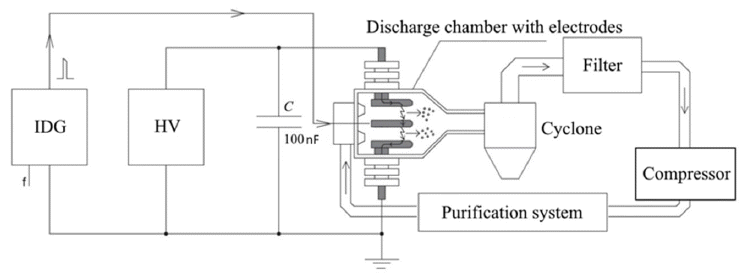
- Nickel nanopowders were obtained using the setup, based on the spark discharge method [5]. The schematic diagram is shown in Fig. 1. The material of all installed electrodes is Ni with a purity of 99.99%. All samples were obtained in a pure N2 atmosphere. Upon completing the operation for 1hr, the collection and conservation of each powder samples were carried out to prevent their oxidation in air. Hexane was used as a conserver.
- A high-voltage source VIP-50 (HV) charged a highvoltage capacitor with a capacitance equal to 100 nF and voltages of 10-17 kV. The gap between the electrodes were varied from 8 to 15 mm. A short high-voltage pulse was applied by the ignition discharge generator (IDG) which ensured ignition of the capacitor discharge through all electrodes at a fixed frequency from 30 to 80 Hz. The frequency was set as to provide a maximum power of HV at fixed voltage. Under the action of erosion by an electric discharge, a cloud of evaporated materials was formed between the electrodes. The nitrogen carrier gas with 40 L/min flow rate supplied by compressor took away the evaporated electrode material from the discharge chamber in which evaporation cloud was subsequently cooled and condensed into nanoparticles. In a cyclone separator (CS), the separation of the fine fraction of nanoparticles from the coarse one has been done. After that, the nanoparticles were accumulated on the filter where the obtained samples were collected.
- The purification system included a mechanical particle filter and an oxygen absorber. Oxygen absorption was provided by heating the titanium sponge up to 700°C. Oxygen control in the chamber of the setup was carried out using an electrochemical oxygen partial pressure sensor (RU 2 395 832 C1).
- The powders were analyzed by the methods of XRD, EDX, TEM and HRTEM. X-ray phase analysis was carried out on the D8 Discover X-ray Diffractometer (Bruker AXS) in copper radiation with a wavelength of 0.154 nm. The diffraction patterns were recorded on copper radiation (Cu Kα, λ=1.542 Å) with a graphite monochromater on a diffracted beam. Processing was performed using the TOPAS 3 program. Electron microscopy was performed on a transmission electron microscope JEOL JEM 2100 at an accelerating voltage of 200 kV.
2. Experimental
- 3.1 XDR analysis
- The XDR analyses results of obtained samples showed that 8-10% of the nickel nanoparticles mass with coherent scattering region (CSR) (19 ± 2 nm) have a cubic structure with a = (0.3529 ± 0.0007 nm), which corresponds to the literature data for a bulk material (PDF No. 004-0850, a = 0.35238 nm). The main crystalline phase (about 90 wt.%) is nickel with a cubic structure with CSR (1.5-2 nm) and a large lattice period a ≈ 0.379 nm (Table 1). One of the possible reasons of this phenomenon may be connected with rapid cooling of nanoparticles during their production.
- When evaluating the average crystallite size (CSR), the correction coefficient K (in the Scherer formula) equal to 0.89 was used.
- Discharge energy was calculated by following equation (1).
- where C is capacitance of the discharge circuit; U is discharge voltage. The energy introduced into a spark was much less than the stored energy and was about 10%, based on our estimates. The rest of the energy was dissipated on the resistance of current lines, gas ionization and electrodes heating.
- XRD showed that the phase does not contain nickel oxide. Based on this, we are able to conclude that the obtained samples during the analysis were metallic.
- 3.2 TEM, HRTEM and EDX analysis
- A small amount of the nanopowder was dispersed in ethanol and then ultrasonically deposited on copper grids containing a carbon film. The samples were placed in a vacuum desiccator to dry at ambient temperature. TEM images and size distribution of Ni nanoparticles was shows in Fig. 2. Most of the large individual Ni particles are predominantly spherical with smooth surfaces and uniform size. Most of the small particles are connected to each other and form chains. These chains are formed as a result of agglomeration of nanoparticles due to surface tension forces and attraction of nanoparticles and due to magnetic forces. Agglomerates are mesoporous spatial 3D nanostructures that consist of aggregates of nanoparticles. In turn, the aggregates are composed of crystalline, non-porous nanoparticles.
- The size distribution is plotted from the TEM images. As can be seen, the largest fraction is particles less than 5 nm. However, there are some large nanoparticles, more than 50 nm. The total number of particles was about 1000. The size distribution results showed that the average nanoparticle diameter was 4.9 nm and the geometric standard deviation (GSD) was 1.26.
- The EDX analyses shown in Fig. 3 were performed on three sample sites showing the absence of possible impurities from elements (N, Ar) in the samples. The samples contained oxygen of 7.54 wt%. (2.17% atomic). From these results, it can be concluded that the nickel nanoparticles are pure. The presence of oxygen can be explained by:
• Oxygen could be adsorbed on the nanoparticle surface during storage.
• Adsorption of oxygen at preparation for microscopy.
- The HRTEM image of 111 crystal plane is shown in Figure 4. The interplanar distance was 0.23 nm. Image processing and analysis was performed using the Gatan Digital Micrograph version 3.9.3. An analysis of the images showed the presence of pure nickel in the sample with a lattice parameter of 0.358 nm, which corresponds to ordinary nickel. In addition, the crystallites contain nickel oxide with lattice parameter of 0.419 nm and nickel with a = b = 0.458 nm, c = 1.299 nm.
- Points (reflexes) in the pictures of the fast Fourier transform (FFT) are blurry. This indicates an uneven interplanar distance.
3. Results and discussion
- Ni nanoparticles synthesized by the spark discharge method have been carried out. XRD results demonstrate the possibility of obtaining metal nanoparticles by spark discharge apparatus. The X-ray diffraction patterns revealed an increase in the lattice period of Ni nanoparticles. This is probably due to the nonequilibrium conditions of the small nanoparticles formation. TEM indicates an irregular crystal lattice period. This may be due to partial oxidation of the sample.
4. Conclusion
- 1. M. Gamarnik: Phys. Status Solidi B, 168 (1991) 389. .Article
- 2. G. Okram, Kh. Devi, H. Sanatombi, A. Soni, V. Ganesan and D. Phase: J. Nanosci. Nanotechnol., 8 (2008) 4127. .ArticlePubMed
- 3. X. Liu, H. Zhang, K. Lu and Z. Hu: J. Phys.: Condens. Matter., 6 (1994) L497. .Article
- 4. R. Birringer and P. Zimmer: Acta Mater., 57 (2009) 1703. .Article
- 5. I. Beketov, A. Bagazeev, E. Azarkevich, A. Maximov, A. Medvedev and A. Beketova: Russian Phys. J., 61 (2018) 166. .
Figure & Data
References
Citations
Citations to this article as recorded by 

- Comparative Study of Spots and Craters Formed during Spark Discharge in Air on Electrodes Made of Different Metals
A. D. Maksimov, E. I. Azarkevich, I. V. Beketov, D. S. Koleukh
Bulletin of the Russian Academy of Sciences: Physics.2025; 89(10): 1941. CrossRef - Comparative Analysis of Craters Formed on Cathode and Anode Spots of a Spark Discharge in Air on Iron Electrodes
A. D. Maksimov, E. I. Azarkevich, I. V. Beketov, D. S. Koleukh
Bulletin of the Russian Academy of Sciences: Physics.2023; 87(S2): S274. CrossRef
Features of Nickel Nanoparticles Structure Synthesized by the Spark Discharge Method


Fig. 1
The scheme of experimental setup.
Fig. 2
TEM images of Ni nanopowder and size distribution.
Fig. 3
Energy-dispersive X-ray spectroscopy of Ni nanopowder sample.
Fig. 4
HRTEM image of Ni nanopowder sample, insert is FFT of selected area.
Fig. 1
Fig. 2
Fig. 3
Fig. 4
Features of Nickel Nanoparticles Structure Synthesized by the Spark Discharge Method
Table 1
Content of the main crystalline phase depending on the experimental setup parameters
Table 1
TOP
 KPMI
KPMI


 Cite this Article


