
Articles
- Page Path
- HOME > J Powder Mater > Volume 30(1); 2023 > Article
-
Article
- Selective Laser Sintering of Co-Cr Alloy Powders and Sintered Products Properties
- Dong-Wan Leea, Minh-Thuyet Nguyena,b, Jin-Chun Kima,*
-
Journal of Korean Powder Metallurgy Institute 2023;30(1):7-12.
DOI: https://doi.org/10.4150/KPMI.2023.30.1.7
Published online: January 31, 2023
a School of Materials Science and Engineering, University of Ulsan, 93 Daehak-ro, Nam-Gu, Ulsan 44610, Republic of Korea
b Hanoi University of Science and Technology, 1 Dai Co Viet, Hai Ba Trung, Hanoi 100000, Vietnam
- * Corresponding Author: Jin-Chun Kim, TEL: +82-52-712-8061, FAX: +82-52-712-8045, E-mail: jckimpml@ulsan.ac.kr
- - D.-W. Lee: 학생, M.-T. Ngyuen: 전임강사, J.-C. Kim: 교수
• Received: January 26, 2023 • Revised: February 16, 2023 • Accepted: February 17, 2023
© The Korean Powder Metallurgy Institute. All rights reserved.
- 2,052 Views
- 27 Download
Abstract
- Metal-additive manufacturing techniques, such as selective laser sintering (SLS), are increasingly utilized for new biomaterials, such as cobalt-chrome (Co-Cr). In this study, Co-Cr gas-atomized powders are used as charge materials for the SLS process. The aim is to understand the consolidation of Co-Cr alloy powder and characterization of samples sintered using SLS under various conditions. The results clearly suggest that besides the matrix phase, the second phase, which is attributed to pores and oxidation particles, is observed in the sintered specimens. The as-built samples exhibit completely different microstructural features compared with the casting or wrought products reported in the literature. The microstructure reveals melt pools, which represent the characteristics of the scanning direction, in particular, or of the SLS conditions, in general. It also exposes extremely fine grain sizes inside the melt pools, resulting in an enhancement in the hardness of the as-built products. Thus, the hardness values of the samples prepared by SLS under all parameter conditions used in this study are evidently higher than those of the casting products.
- The Co-Cr alloys have been got great attention from many researches and manufactures due to their remarkable properties such as high resistance to wear and corrosion. Co-Cr alloys also have a broad range of applications in making wind turbines, engine components and many other industrial applications where high wear resistance is needed. In addition, Co-Cr alloys are known as important materials in the biomedical industry. They are commonly used to make artificial joints including knee and hip joints [1, 2], or for removable partial dentures, metal frames due to high wear-resistance and biocompatibility. Up to now, Co-Cr alloys have been used in both types of the cast and wrought products. However, the conventional processes for fabrication these alloys, such as casting, cutting, and plastic works, are usually the long-time processes and also difficult because of the natural reasons of high melting point, hardness, and limited ductility [3-6]. In order to improve the quality and the efficiency of products, load of methods have been investigated and applied up to now. Among advantaged manufacturing techniques, additive manufacturing (AM) exhibited a high potential not only in term of reducing production time due to its capability to directly build up threedimensional parts with complex shapes, but also in term of bring out the high quality for the final products [7, 8]. Therefore, AM have become promising techniques for fabricating the medical products in general and biocompatible Co-based alloys in particular [6, 9, 10]. In recent years, AM processes in which a laser beam was used to selectively melt powder materials such as selective laser melting (SLM) and selective laser sintering (SLS) have been widely applied. In SLM/SLS, metal powders are irradiated by a laser beam with spot size in micro scale, therefore a micro melt pool could be formed, leading to a high cooling rate (103 ~ 108 K/s) [11]. A complex heat effect acts on a material, resulting in a different heat transfer mechanism compared with that in casting and forging. 316L stainless steels [12], Ni625 [13], AlSi10Mg [14], Ti-6Al-4V [9] and Co-Cr alloys are the most used and studied materials for SLM/SLS techniques [7].
- In case of Co-Cr alloys, most of the studies were conducted on the samples which were produced primarily by SLM process. In which, several particular processing parameters, including scanning strategy, hatch distance, spot size and power of the laser have been used in order to investigate the microstructure and mechanical properties of as-built Co-Cr samples and the difference between them with conventional products [15, 16]. There are few studies reported about the using of SLS to produce Co-Cr products, therefore, there is a lot of rooms for filling up in this field. For examples, SLS processing parameters must be optimized, the mechanism of defects, pores and microstructure formations need to be figured out in order to understand the effects of SLS processing on structural components and to optimize the parameters in order to obtain the higher quality of products.
- In this study, a SLS process was used to manufacture Co-Cr specimens from powder type. The investigation was deeply conducted in order to recognize a better understanding of their typical microstructure, microstructural defects on the as-built samples and their hardness behavior. In particular, specimens built with various parameters were used to investigate the effect of conditions on as-built samples properties and determine the optimized processing parameters, which is useful reference for future researches or applications. Finally, the effect of microstructure according to process variables on the hardness of the specimen was analyzed.
1. Introduction
- 2.1. Material information and sintering parameters
- Gas-atomized Co-Cr alloy powders with the chemical composition (in wt.%) and particle size was present in Table 1 were used in this study.
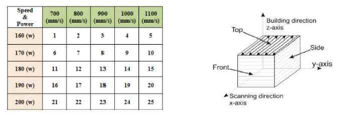
- The 3D dimensions specimens were prepared by a selective laser sintering system which was operated with the various parameters as showed in Table 2. In this study, 25 cubic samples with dimensions of 10 mm × 10 mm × 10 mm were built under Ni atmosphere. For easily in determination, the as-built specimens were numbered from 1 to 25 as presented in Fig. 1(a).
- During SLS process, a zigzag scanning strategy was used. Furthermore, in order to minimize anisotropy and to obtain the dense of products, each layer was built with the laser scanning along a specific direction. In addition, layer-by-layer the scanning direction was rotated by 67- 690 in comparison to the previous one, therefore, the same pattern is repeated every 180 layers. The scanning direction for the last layer on top of final products is illustrated in Fig. 1(b).
- 2.2. Phase and Microstructural characterization
- X-ray diffraction (XRD) was used to determine the phase structures of samples. XRD measurements were performed with a Cu-Kα radiation source at V = 40 kV and I = 40 mA in the angular range 2θ = 20-100°. Microstructural characterizations of the as-built specimens were carried out by optical microscopy (OM) and scanning electron microscopy (SEM) techniques. The analyses were performed on cross-sectioned sintered samples. Before observations, as-built specimens were ground and polished by using a conventional metallographic procedure and chemical etching in the HF solution for about 20 seconds.
- 2.3. Hardness measurements
- Microhardness tests were conducted on the sintered samples using the micro-Vickers hardness (Mitutoyo MVK-H1) with the load of 500 g. Measurements were obtained averaging five indentations for each specimen.
2. Experimental


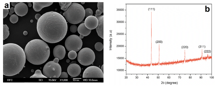
- Morphology and XRD pattern of powder are shown in Fig. 2. From the SEM result (Fig. 2(a)), the shapes and the average size of powders were evaluated. The powder exposes the very spherical shape, smooth surface and the size of the particles ranges from 5 to 40 μm which is suitable for SLS process [17]. Fig. 2(b) displays the XRD pattern of the alloy powders. According to the XRD profile, all the visible peaks can be attributed to the cubic cobalt phase demonstrated that this alloy exhibits a typical FCC crystal structure, and in general those peaks corresponded to as γ phase which is in close agreement with the results reported in the literature of Co-Cr alloys [18].
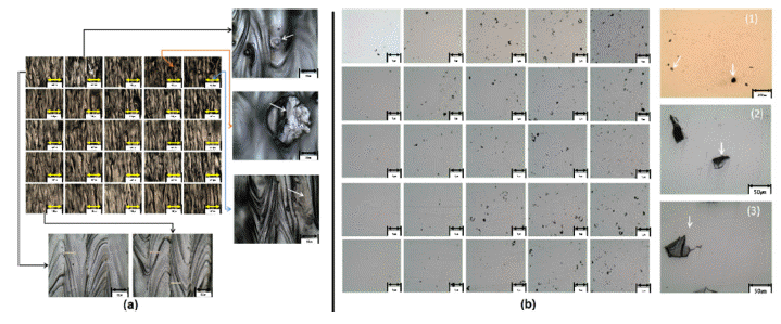
- Optical microscopy observations were performed on the surface of the sintered samples right after sintering and after polishing with a suitable technique and the results are shown in Fig. 3. The order of samples matches with the arrangement in Fig. 1(a). For the as-built samples before polishing (Fig. 3(a)), the top surface lines that characterize the laser scanning strategy of zigzag type and the hatch spacing of 70 μm could be measured. It also reveals that there were still un-sintered powders could be seen on the surface of the as-built samples right after laser scanning took place, especially when it was conducted with low laser power (Fig. 3(a)).
- In polishing condition (Fig. 3(b)), it can be seen that the rough surface has been removed and the internal phase of the specimens were lightly revealed. The OM images show that there are two phases were observed matrix phase (bright area) and the second dark phase (dark regions). The second phases here are attributed to the laser traces (3), vacancies (2) or oxidation phases (1) which are presented in the high magnification images in Fig. 3(b). The optical images of the surface sintered samples right after polishing also give us more detail about the quality of SLS process. It could be seen that with each laser power parameter, the number of the second phases slightly increased with the increase in scanning speed. However, the number and the distribution of the second phases showed the same and were not strongly affected by the laser power even at high laser scanning speeds. Contrarily, with low speed (700-800 mm/s) of scanning the as-built samples exhibited smoother surfaces and lesser of the second phase at higher laser powers. From those results, we could asserted that it was better to conduct SLS process at low scanning speed, especially around 700-800 mm/s and high laser power from 180-200 (W).
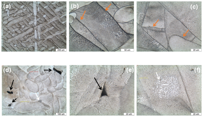
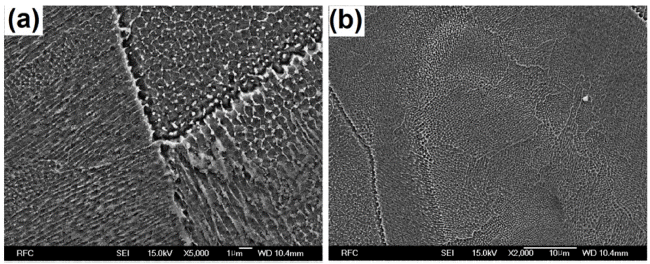
- After etching with a suitable solution, the etched asbuilt Co-Cr samples exposed their structure visibly as presented in Fig. 4. According to those images, the asbuilt products showed the totally different microstructure in comparing with casting [1, 19, 20], spark plasma sintering [4] and wrought Co-Cr alloys [3, 5] in literature. The most difference features could be clearly recognized between them were the absence of dendritic structure and carbide phase which were typical properties of the cast Co-Cr alloys in SLS samples in this study. Additionally, their structure images implied the characteristics reflecting the properties of process parameters which could not be recognized in any conventional products. In particular, as we can see in Fig. 4(a), the melt pools and their directions characterized the laser scanning strategy and laser directions. The layer-by-layer the scanning direction and the rotation of layer after layer were also revealed clearly. It is worth noting that the laser scanning direction was rotated by 67-69° to previous layer, leading to differently oriented melt pools.. The lines separating the different weld pools produced by the laser scanning on each layer are evidenced obviously by arrows in Fig. 4(b) and Fig. 4(c). Furthermore, the dimensions of each melt pool were proximately 70-80 μm in width, a bit larger than the hatch spacing of 70 μm caused by the expansion of melted zone. Therefore, we definitely confirmed that these features reflected the SLS parameter conditions.
- The higher magnification images of the inner structure of the sintered specimens demonstrate the presence of an extremely fine microstructure inside a single pool. Fig. 4 (b), (c) exposes the typical columnar structures grew inside the matrix with the direction perpendicular to the melt pool boundaries, which is also the directions of the highest temperature gradient. It could be seen more clearly in Fig. 5(a). At the center of the melt pools, there were many domains of very tiny columnar structure which was grown in different directions, and the primary cell spacing of the structure was approximately 1 μm, Fig. 5(b). Up to now, the cause of the formation that kind of microstructure in as-built Co-Cr specimens has been attributed to the melting and solidification dramatically after powder had been scanned by the laser. However, it needs more studies to figure out a concrete mechanism.
- Observations performed on the etched surface of asbuilt samples allow evidencing the presence of the defects and the second phases in the Co-Cr matrix. They were pores (Fig. 4(e)), laser traces or oxidation phases as could be seen in Fig. 4(d) and the microstructure of powder region (Fig. 4(f)). The presence of pores and laser traces were attributed to the lack of fusion during scanning process or caused by the trapped gas bubbles in the initial materials [13]. Besides pores, microcracks also exist in some samples (Fig. 4(e)), were due to the shrinkage of material when the powder was melted by the effects of laser and then cooled down rapidly. Fig. 4(f) shows an area with the microstructure seem like microstructure of the initial powders indicating that there was few un-melted powder particles but it welded well with melted parts to make a uniform matrix in as-built samples. In SLS process, laser beam scanned on powder layer, then, powders were melted and consolidated rapidly. Therefore, products were built in one step without any refining or removing of impurities phases as in conventional melting process resulting in the existence of the small amount of slag known as oxidation phases in the final samples.
- The microhardness values of as-built samples are shown in Table 3. It shows a great variation in Vickers microhardness. However, most of them are above 430 Hv. Which is very high value in comparison to the cast Co-Cr alloys (~300 Hv) and even higher than SPS Co-Cr alloys (max.415 Hv) [3, 4]. The better hardness properties of as-built samples could be explained by the Hall- Petch effects when a very finer grain size was obtained..
3. Results and discussion
- Co-Cr alloy sintered samples were successfully built by using SLS technique with different parameters of laser speed and laser power. There were 2 phases including matrix (white) and the second phases (dark) which is attributed to vacancies, laser traces and oxidation existed in as-built products. The microstructure of samples represents the characteristics phases in the manufacturing process. It is not similar to the dendritic phases in casting but the solidification morphologies of the laser beam melting. The SLS process allowed obtaining samples with improved chemical homogeneity over the molten sample and an extremely tiny structure and a refined grain, originated by the rapid and localized solidification, resulting in great mechanical properties could be achieved. We investigated the effect of conditions on asbuilt samples properties and determine the optimized processing parameters. This study also indicated that it was better when conducted SLS at low speed to reduce defects in the sintered samples.
4. Conclusions
-
Acknowledgements
- This results was supported by “Regional Innovation Strategy (RIS)” through the National Research Foundation of Korea (NRF) funded by the Ministry of Education (MOE) (2021RIS-003).
Acknowledgments
- 1. J. A. Pieniazek, A. Lukaszczyk and R. Zapala: Arch. Metall. Mater., 58 (2013) 1281..Article
- 2. D. J. S. Hyslop, A. M. Abdelkader, A. Cox and D. J. Fray: Acta Mater., 58 (2010) 3124..Article
- 3. S.-H. Lee, E. Takahashi, N. Nomura and A. Chiba: Mater. Trans., 46 (2005) 1790..Article
- 4. N. Vicentea, F.Casari, F. Bucciottic, L. Facchinic and A. Molinaria: Euro PM2011–PM Biomaterials, (2011)..
- 5. A. L. R. Ledesma, H. F. Lopez and J. A. J. Islas: Metals, 6 (2016) 188..Article
- 6. A. Takaichi, Suyalatu, T. Nakamoto, N. Joko, N. Nomura, Y. Tsutsumi, S. Migita, H. Doi, S. Kurosu, A. Chiba, N. Wakabayashi, Y. Igarashi and T. Hanawa: J. Mech. Behav. Biomed. Mater., 21 (2013) 67..ArticlePubMed
- 7. W. E. Frazier: J. Mater. Eng. Perform., 23 (2014) 1917..Article
- 8. S. Mellor, L. Hao and D. Zhang: Int . J. Prod. Econ., 149 (2014) 194..Article
- 9. L.-C. Zhang and H. Attar: Adv. Eng. Mater., 18 (2016) 463..Article
- 10. B. Vandenbroucke and J.-P. Kruth: Rapid Prototyping J., 13 (2007) 196..Article
- 11. W. Shifeng, L. Shuai, W. Qingsong, C. Yan, Z. Sheng and S. Yusheng: J. Mater. Process. Technol., 214 (2014) 2660..Article
- 12. R. Casati, J. Lemke and M. Vedani: J. Mater. Sci. Technol., 32 (2016) 738..Article
- 13. C. Li, R. White, X. Y. Fang, M. Weaver and Y. B. Guo: Mater. Sci. Eng. A, 705 (2017) 20..Article
- 14. N. Read,, W. Wang, K. Essa and M. M. Attallah: Mater. Des., 65 (2015) 417..Article
- 15. Y. Kajima, A. Takaichi, T. Nakamoto, T. Kimura, Y. Yogo, M. Ashida, H. Doi, N. Nomura, H. Takahashi, T. Hanawa and N. Wakabayashi: J. Mech. Behav. Biomed. Mater., 59 (2016) 446..ArticlePubMed
- 16. E.Girardin, G. Barucca, P. Mengucci, F. Fiori, E. Bassoli, A. Gatto, L. Iuliano and B. Rutkowski: Mater. Today Proc., 3 ( 2016 ) 889..Article
- 17. J. A. Slotwinski, E. J. Garboczi, P. Stutzman, C. F. Ferraris, S. Watsoon and M. Peltz: J. Res. Natl. Inst. Stand. Technol., 119 (2014) 460..ArticlePubMedPMC
- 18. A. J. Saldivar-Garcia and H. F. Lopez: Metall. Mater. Trans. A, 35 (2004) 2517..Article
- 19. H. R. Kim, S.-H. Jang, Y. K. Kim, J. S. Son, B. K. Min, K.-H. Kim and Y.-T. Kwon: Materials, 9 (2016) 596..ArticlePubMedPMC
- 20. J. V. Giacchi, C. N. Morando, O. Fornaro and H. A. Palacio: Mater. Charact., 62 (2011) 53..Article
Figure & Data
References
Citations
Citations to this article as recorded by 
 Cite this Article


Selective Laser Sintering of Co-Cr Alloy Powders and Sintered Products Properties


Fig. 1
(a) Numbering of specimen versus power and laser speed. (b) Scanning and building direction.
Fig. 2
Morphology and XRD patterns of the as-received Co-Cr alloy powder.
Fig. 3
Optical microscopy images of the as-built specimens: (a) - before and (b) - after polishing.
Fig. 4
As-built microstructure of Co-Cr samples after etching in a suitable solution.
Fig. 5
Structures of as-built sample at area (a) near boundary and (b) center of melt pool.
Fig. 1
Fig. 2
Fig. 3
Fig. 4
Fig. 5
Selective Laser Sintering of Co-Cr Alloy Powders and Sintered Products Properties
Table 1
Composition and particle size analysis of the as-received powder
Table 2
The parameter condition used in SLS process
Table 3
Microhardness values of as-built samples (Hv)
Table 1
Table 2
Table 3
TOP
